
Neuroendoscopy
 Hydrocephaly is a disease appearing when the cerebrospinal fluid outflow from the brain ventricles (inner reservoirs) is blocked. A ventriculoperitoneal shunting is traditionally used to take excess fluid from the ventricles. A thin tube is inserted into a ventricle and then it passes through a valve into the abdominal cavity or large vein.
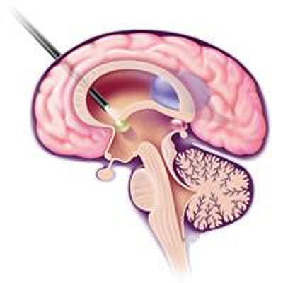
In the 90s of the XX century a revolutionary method for perforation of the third ventricle’s bottom is offered – neuroendoscopic. The neuroendoscope was invented on the basis of the last technical achievements of the humankind. This device is actually a complicated optical system of the Hopkins’ lenses, a lighting channel, a videochannel, as well as the channels for infusion and averting the fluid, embodied in the tube only 6 mm in diameter. The operation is performed through the manipulation channel. The picture is transmitted on the screen and the doctors see where they should direct their instruments, so that the outflow of the liquid could be renewed.  The surgery aims at creating the outflow via the third ventricle’s bottom to the out-brain cisterns of the liquor system. A new opening is created by a special catheter. As a result, the neurolymph flows out of the brain without any obstruction, and the person is saved forever.
This method is low traumatic and reliable. Unlike the shunt which takes an hour or two, this operation lasts only 10-20 minutes.
In general, inserting and manipulating the neuroendoscope require sufficient skills and perfect handling of the equipment.
The doctors of the International Neurosurgery Center learnt Neuroendoscopy in 2004 from Henry Marsh, the English neurosurgeon who was the pioneer in using this method in Great Britain (1994).
It is possible to use the neuroendoscope for diagnostics and treatment of some brain tumors and cysts.